
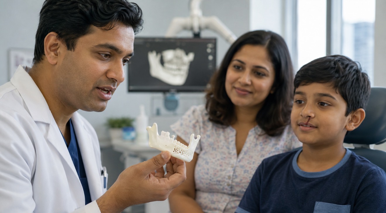
A new biodegradable material developed by Australian researchers could eventually change the way children with cleft lip and palate undergo one of the most difficult stages of their treatment.
Researchers at the University of Sydney have developed what they describe as a “nanobone”, a calcium-based nanomaterial designed to encourage the body to regenerate its own bone rather than relying entirely on bone taken surgically from another part of the patient’s body.
In preclinical testing, the material generated approximately 80 percent more new bone than the control material after eight weeks and activated a naturally occurring bone-repair growth factor at levels around 10 times higher than conventional methods, according to the research team.
The work, led by Associate Professor Chun Xu from the University of Sydney’s Faculty of Medicine and Health, has been published in the scientific journal ACS Nano.
The technology remains experimental and has not yet been tested in human patients.
But if further studies and clinical trials eventually confirm its safety and effectiveness, researchers believe it could offer a less invasive approach to repairing difficult bone defects, particularly for children born with cleft lip and palate.
Why bone repair is such a difficult part of cleft treatment
Cleft lip and cleft palate occur when structures that form the upper lip or roof of the mouth do not completely join during pregnancy.
The condition affects roughly one in every 700 children internationally.
Many of the visible aspects of a cleft can be treated during infancy.
But when the cleft extends through the gum and jawbone, another significant procedure is often required years later.
Children with an alveolar cleft commonly undergo bone graft surgery when they are around nine to 12 years old, before certain permanent teeth emerge.
The procedure usually involves removing bone from another part of the child’s body, often the hip, and transferring it into the gap in the upper jaw.
That means surgeons are effectively creating a second surgical site simply to obtain the material required to repair the first.
Associate Professor Xu said that delay and additional surgery can create a considerable burden for children and families.
“One of the biggest challenges for children born with cleft lip and palate is repairing the bone defect in the jaw,” he said.
While some smaller defects can be addressed earlier, many children must wait until later childhood before undergoing grafting.
During those years, the condition can affect eating, breathing, speech, dental development and confidence.
What exactly is ‘nanobone’?
The term sounds futuristic, but the concept behind it is relatively straightforward.
Instead of supplying the body with externally manufactured growth factors, the new material is designed to activate repair mechanisms that are already present naturally.
The material is made from calcium-aluminosilicate nanoparticles.
Researchers found it could activate a naturally occurring protein known as latent Transforming Growth Factor beta-1, or TGF-β1.
TGF-β1 plays an important role in tissue repair.
Once activated, it helps attract bone-forming stem cells towards the damaged area and encourages those cells to develop into cells capable of producing new bone.
The ultimate objective is therefore not for the artificial material to permanently replace the missing bone.
The material acts as a catalyst for healing.
As regeneration progresses, the material is intended to degrade while the patient’s own bone tissue takes its place.
Four jobs from one material
One particularly interesting aspect of the Sydney research is that the material appears capable of performing several functions involved in bone repair.
|
Function |
What researchers observed |
|
Blood clotting |
Helped promote clot formation in around 30 seconds |
|
Growth-factor activation |
Activated the body’s own latent TGF-β1 |
|
Stem-cell recruitment |
Encouraged bone-forming cells towards the damaged site |
|
Bone regeneration |
Produced substantially greater new bone formation in the preclinical model |
|
Long-term role |
Designed to biodegrade as natural tissue replaces it |
The researchers say this is the first demonstration of a single nanomaterial platform combining rapid clotting, activation of endogenous growth factors, recruitment of bone-forming stem cells and enhanced bone regeneration.
That combination is important because successful bone repair involves much more than filling a physical hole.
A damaged area has to stabilise.
Blood supply needs to develop.
Repair signals have to be activated.
Cells capable of creating bone must reach the area.
Those cells then have to produce structurally useful tissue.
The body already has much of the machinery required
Xu said the research was based on the idea that the human body already possesses many of the biological signals required for tissue repair.
“Our body already contains many of the signals needed for tissue repair,” he said.
“We’ve developed a material that can help activate those signals at the right place and time.”
That represents a different approach from treatments that supply manufactured biological growth factors externally.
Rather than attempting to replace the body’s repair system, the Sydney team is trying to activate it.
Why the 80 percent result matters, and why caution is still needed
The headline laboratory result is significant.
In the preclinical bone model used by researchers, the nanomaterial produced approximately 80 percent more new bone than the comparison material after eight weeks.
Researchers also reported roughly 10 times the level of activation of the targeted bone-repair growth factor compared with conventional approaches.
Those findings justify further investigation.
They do not, however, mean children can expect to receive this treatment soon.
The research remains at the preclinical stage.
That distinction is crucial.
Many technologies demonstrate impressive results in laboratory or animal models but face additional challenges when eventually tested in people.
Researchers will still need to examine issues including:
- long-term safety
- degradation of the material
- inflammatory responses
- optimal dosage
- consistency of bone growth
- effectiveness in different types and sizes of defects
- interaction with growing children’s bones
- manufacturing and regulatory requirements
Human clinical trials would only follow if earlier stages continue to produce convincing safety and efficacy results.
Researchers hope treatment could eventually happen earlier
Perhaps the most important long-term ambition is not simply replacing one material with another.
It is potentially changing when treatment can happen.
Traditional bone grafting for cleft-related jaw defects is often timed around the development of permanent canine teeth.
Researchers hope a regenerative material might eventually allow doctors to intervene earlier in childhood.
That could potentially shorten the period during which a child lives with an unrepaired jaw defect.
Xu said the team’s long-term goal was to develop materials that allow the body to regenerate bone naturally while reducing reliance on invasive and painful procedures.
If that ultimately proves possible, it could change a treatment pathway that has remained broadly similar for decades.
The technology could go beyond cleft lip and palate
The research also has implications outside paediatric cleft treatment.
More than four million bone-repair procedures are performed around the world each year, according to the University of Sydney team.
Bone defects can result from:
- traumatic injuries
- dental disease and tooth loss
- congenital conditions
- surgery
- infection
- cancer treatment
- complex fractures
Autologous bone grafting, where bone is taken from another part of the same patient, remains an important treatment because the material is biologically compatible.
But removing that bone can mean additional pain, recovery time and surgical complications.
A material capable of safely stimulating natural bone regeneration could therefore have applications across dentistry, orthopaedic surgery and reconstructive medicine.
3D printing could take the idea another step
The Sydney researchers are also studying how nanobone could be combined with 3D-printed scaffolds.
This could potentially allow surgeons to create structures designed around the exact dimensions of an individual patient’s bone defect.
That matters because no two injuries or congenital defects are identical.
Xu said the longer-term goal was personalised treatment.
“Every patient is different and every bone defect is different,” he said.
“In the future, we hope to combine these materials with advanced 3D-printing technologies so treatments can be tailored to the specific needs of each patient.”
In principle, medical imaging could map the shape of a defect, a scaffold could then be printed to fit that particular area, and regenerative material could help encourage the patient’s own tissue to grow through it.
That remains a future possibility rather than established clinical practice.
But it demonstrates where regenerative medicine is heading.
Why New Zealand should be watching this research closely
Although the discovery comes from Australia, its relevance does not stop at the Tasman Sea.
New Zealand children undergo the same long and often complex treatment journey after being born with cleft lip or palate.
Cleft New Zealand estimates that approximately one in every 700 babies is born with a cleft lip and/or palate.
New Zealand recorded 56,268 live births in the year ended June 2026. At a prevalence of around one in 700, that would equate very roughly to about 80 babies in a typical year, although the actual number will vary from year to year.
That means this is not an obscure condition affecting only a handful of families overseas.
There are New Zealand families beginning this treatment journey every year.
New Zealand children also face bone grafting around the same age
Current information provided by Cleft New Zealand describes a treatment pathway remarkably similar to the one highlighted by the Australian researchers.
For children whose cleft extends through the gum area, bone grafting is typically carried out when permanent canine teeth are developing, often somewhere between the ages of nine and 12.
Bone may be taken from another part of the child’s body, such as the hip, and placed into the gap in the jaw.
Orthodontic treatment is also usually required before the graft procedure to prepare the site.
For New Zealand families going through that process, research that could eventually reduce the need for a second surgical site is therefore highly relevant.
The real benefit could be reducing the burden on children
It is easy to look at this research primarily as a technological achievement.
But the more important question is what it could mean for a child.
Cleft treatment can stretch across much of childhood.
There can be surgery in infancy.
Speech therapy.
Dental appointments.
Orthodontics.
Bone grafting.
And, for some young people, additional jaw or cosmetic surgery later.
Each procedure also means hospital visits, recovery, school disruption and anxiety for families.
A technology that safely eliminates even one major invasive procedure could therefore have an impact extending well beyond the operating theatre.
Australia and New Zealand should collaborate as the research develops
There is also a strong argument for closer trans-Tasman cooperation if the technology progresses towards clinical testing.
Australia has larger research institutions and patient populations.
New Zealand has established multidisciplinary cleft services and families who could ultimately benefit from advances in treatment.
Both countries operate comparable health and medical regulatory environments and already share substantial research connections.
If the nanobone technology progresses successfully through further preclinical research and into human trials, New Zealand clinicians and researchers should be watching closely.
Participation in future collaborative studies may also be worth considering if appropriate clinical and ethical requirements can be met.
A promising discovery, not yet a medical breakthrough
The most responsible way to describe this research is with cautious optimism.
The early results are encouraging.
The biological mechanism is interesting.
The potential clinical need is genuine.
And the possibility of encouraging a child’s body to repair its own jawbone rather than harvesting bone elsewhere is compelling.
But this is not yet an alternative parents can request from their surgeon.
There are still substantial scientific and regulatory steps between a successful preclinical experiment and routine treatment in a children’s hospital.
That process could take years.
For families affected by cleft lip and palate, however, the research offers something important: evidence that a procedure that has remained broadly unchanged for decades may not always have to be done the same way.
If researchers can eventually turn those early results into safe human treatment, the biggest achievement will not be the nanoparticles themselves.
It will be giving a child’s own body a better opportunity to rebuild what is missing.
Research: Chun Xu, Q. Chen, H. Chen and colleagues, “Multifunctional Mesoporous Calcium-aluminosilicate Nanoparticles Enable Heterogeneous Activation of Endogenous Transforming Growth Factor β1 and Rapid Hemostasis for Bone Repair”, ACS Nano, 2026.
Source: University of Sydney, Faculty of Medicine and Health, School of Dentistry, Charles Perkins Centre and Sydney Nano, in collaboration with the University of Queensland.
New Zealand context: Cleft New Zealand and Stats NZ.